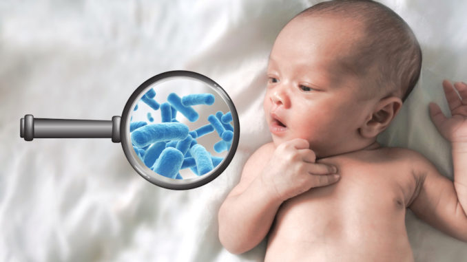
In the largest clinical microbiome study in infants reported to date, a team led by researchers at Baylor College of Medicine explored the sequence of microbial colonization in the infant gut through age 4 and found distinct stages of development in the microbiome that were associated with early life exposures.
Published in the journal Nature, their report and an accompanying report led by the Broad Institute are the result of extensive analysis of data collected from a cohort of participants involved in the TEDDY diabetes study.
The TEDDY study (The Environmental Determinants of Diabetes in the Young) study has been collecting data for 10 years with the goal of understanding what triggers type 1 diabetes in children at increased genetic risk for the disease. Researchers at six clinical centers in the U.S., Sweden, Finland, and Germany, as well as the Data Coordinating Center at the University of South Florida, have gathered monthly stool samples and data from more than 8,600 children who are genetically susceptible to type 1 diabetes. From this cohort, researchers at Baylor College of Medicine analyzed 12,005 stool samples that were collected from 903 children between three and 46 months of age to further understand what the microbiome looks like early in life.
“We know that the first few years of life are important for microbiome establishment. You are born with very few microbes, and microbial communities assemble on and in your body through those first years of your life,”
said Dr. Joseph Petrosino, director of the Alkek Center for Metagenomics and Microbiome Research and professor and interim chair of molecular virology and microbiology at Baylor.
“In this study, we took a closer look in this amazing cohort at the establishment of the microbiome over the first few years of life and the early life exposures associated with that sequence of events.”
Using state of the art sequencing of both RNA and DNA to uncover the complete genetic set up of all microbes, Petrosino and his team determined that the developing gut microbiome undergoes three distinct phases of microbiome progression:
- Developmental phase (3 to 14 months of age)
- Transitional phase (15 to 30 months of age) and
- Stable phase (31 to 46 months of age)
“This information is useful for any future microbiome studies looking at an infant cohort for scientific discovery and potential intervention purposes. The idea that we can stratify the development phases in this manner may give researchers additional resolution to reveal differences that could potentially be disease-associated,” Petrosino said.
More insights into microbiome development
The study found an association between at least partial breastfeeding and having a higher abundance of Bifidobacterium breve and Bifidobacterium bifidum, two types of bacterial species with probiotic properties known to be prevalent early in life. In addition, the cessation of breastfeeding accelerated the maturation of the infant’s microbiome, meaning it proceeded quickly through the other stages to the stable phase, which is hallmarked by higher amounts of the bacteria Firmicutes spp.
“Further research will help better understand the implications of having an accelerated rate of microbiome maturation,”
Petrosino said.
In those infants who were breastfed, the strains of Bifidobacterium that had the genetic capability of processing human milk were no longer detected once breastfeeding stopped.
“The presumption is that selective pressure for these organisms to be present during breastfeeding is removed once breastfeeding stops, and other strains of Bifidobacterium that do not process the metabolites in breast milk can then grow,” Petrosino said. “This provides insight into how the early diet is impacting microbiome development.”
The researchers also found an association between vaginal delivery and having a greater abundance of bacteria belonging to the Bacteroides genus. However, having more Bacteroides at birth was not exclusive to those infants who were delivered by this mode. Those who did have more Bacteroides at birth tended to have a greater diversity of microbes early in the first 40 months of life.
“Again, the implications are not yet clear. Having microbial diversity is typically thought of as beneficial, but we still don’t fully understand which microbial signals early in life are important for development,” Petrosino said.
Petrosino noted that these data already are being used, along with the extensive TEDDY metadata repository, to better understand how environmental exposures contribute to progression to type 1 diabetes. Additional provocative microbiome analyses, including the viral and fungal microbiome constituents, are underway and will also include human genomic, metabolomic and proteomic data, as well as dietary and infectious episode information.
“These initial analyses have reinforced previous infant studies and also have revealed additional important microbiome associations during this critical time in life. Future discoveries from this cohort will pave the way for focused mechanistic work to elucidate how the microbiome influences health and disease, particularly type 1 diabetes,”
said Dr. Christopher Stewart, co-first author of the study, formerly a postdoctoral researcher at the Petrosino lab at Baylor and now a research fellow at Newcastle University.
“It is cohorts such as this, where we can integrate clinical data with patient-specific exposure, genomic and microbiome analyses, that will lead to precision medicine-based diagnostics and therapeutics for type 1 diabetes and many other diseases,”
Petrosino concluded.

