Patients with cystic fibrosis (CF), a genetic disease that causes a persistent buildup of thick mucus in their lungs and other organs, suffer from chronic bacterial infections and inflammation that often lead to deadly respiratory failure. Little is known about how bacteria colonize the lungs or how CF patients respond to these pathogens because there currently is no way to model this process in the laboratory for research and drug development.
A new grant from the Cystic Fibrosis Foundation (CFF) to the Wyss Institute at Harvard University aims to change that by funding the development of a human Airway Chip to recapitulate many of the features of CF lung using the Wyss’ Organ Chip technology.
“This tool will enable researchers to analyze the molecular mechanisms that underlie the lung’s response to pathogens in patients with CF, and provide a human-relevant model in which to test potential CF therapeutics,”
said Wyss Founding Director Donald Ingber, M.D., Ph.D., who is also the Judah Folkman Professor of Vascular Biology at HMS and the Vascular Biology Program at Boston Children’s Hospital, as well as Professor of Bioengineering at Harvard’s John A. Paulson School of Engineering and Applied Sciences (SEAS).
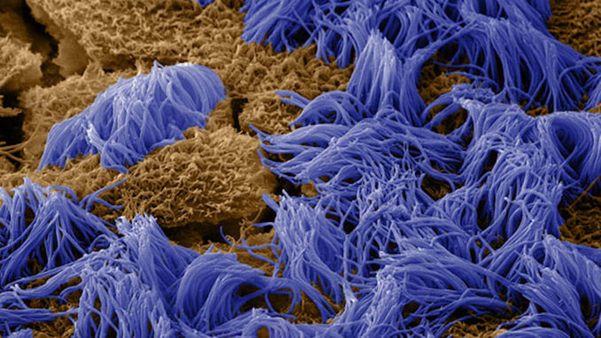
CF, caused by a recessive genetic mutation in the CFTR gene, affects as many as one in every 3,400 newborns of northern European descent in the US and dramatically shortens patients’ lifespans, largely due to chronic respiratory complications (the median survival age is about 40). The CFTR protein normally functions as an ion channel, helping to maintain a thin layer of water on the inner surface of the lung that allows tiny hairs called “cilia” on the outside of the lung cells to sweep back and forth, clearing mucus out of the airways. When the function of the CFTR protein is disrupted, this water layer is broken down, preventing the cilia from clearing mucus and creating a breeding ground for bacteria. The most common pathogen that affects people with CF is the bacterium Pseudomonas aeruginosa, which is usually benign in non-CF patients’ lungs but can become highly infectious due to changes in the CF lung microenvironment. P. aeruginosa is especially dangerous because it can become resistant to attacks by the body’s immune system.
“Organ Chips allow us to create experimental conditions – including co-culturing human cells with living microbes – within an in vitro model that behaves like a human organ, making them the perfect vehicle for studying the ways that bacterial pathogens interact with their hosts at the cellular level,”
said Rachelle Prantil-Baun, Ph.D., a Senior Staff Scientist at the Wyss Institute who is helping Dr. Ingber lead the CF Airway Chip project.
The Wyss Institute has already developed Organ Chips that mimic various organs in the human body, including the liver, kidney, brain, and gut, as well as lung, all of which display human organ-level functions.
“Because all of the parameters in the Organ Chip system can be varied individually, it offers a novel ‘synthetic biology’ approach to study how cellular, molecular, chemical, and physical cues work alone and in combination, to influence human tissue development and disease,”
said Ratnakar Potla, MBBS, Ph.D., a Postdoctoral Fellow at the Wyss Institute who is the Technical Lead of the CF Airway Chip project.
This project will use a modified version of the Wyss’ Lung Small Airway Chip, which was previously used to create a model of chronic obstructive pulmonary disorder (COPD) that successfully mimicked bacterial and viral infections. The research team plans to line the chip’s epithelial channel with CFTR-deficient human airway cells from CF patients and its endothelial channel with healthy blood vessel cells (both provided by the CFF), and analyze the function of those organ models both with and without P. aeruginosa and circulating human neutrophil cells (which mediate inflammation). Because the mutated CFTR gene disrupts the normal flow of ions across the lung epithelium, the CF Airway Chips will also have integrated electrodes to quantify the movement of ions through the CFTR protein channels in real time.
“Once we have this model in place, we will work with the Foundation to prioritize questions that are most relevant to the Foundation and the cystic fibrosis patient community. We believe that through these studies, we will gain a more mechanistic understanding of how pathogens influence cystic fibrosis airway structure and function. We also hope to identify new ways to target the lung to enhance tolerance to relevant pathogens, and possibly novel ways to treat chronic inflammation in these patients,”
said Olivier Henry, Ph.D., a Staff Engineer at the Wyss Institute who is the Sensor Lead of the CF Airway Chip project.
To learn more about cystic fibrosis or the Cystic Fibrosis Foundation, please visit www.cff.org.
